

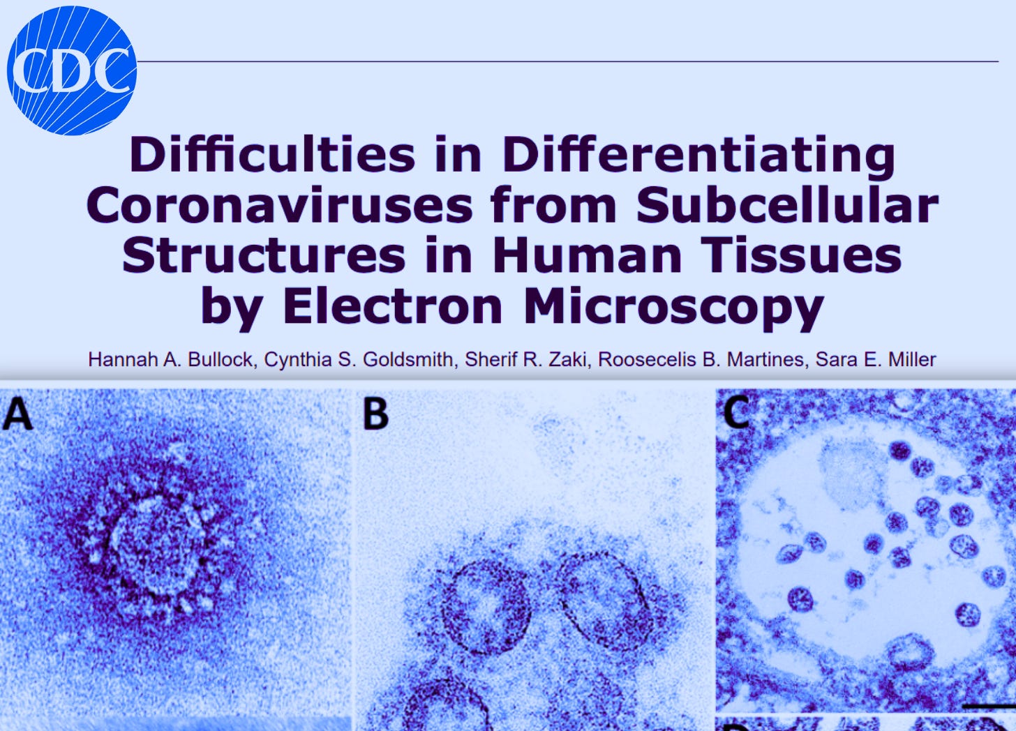
Top medical journals published images of ordinary human cellular structures labeled as “coronavirus,” according to a 2021 review authored by Centers for Disease Control and Prevention (CDC) scientists and collaborators titled “Difficulties in Differentiating Coronaviruses from Subcellular Structures in Human Tissues by Electron Microscopy.”
The researchers examined 27 reports that used electron microscopy to identify coronavirus directly in human tissue and found that 23 contained cellular structures misidentified as virus.
“In each case of erroneously identified coronavirus particles,” they wrote, “the structures mistaken for virus are common cellular organelles.”
More than five years later, several of the papers remain published without corrections withdrawing the disputed electron-microscope identifications, including studies claiming direct SARS-CoV-2 infection of the kidney, liver, heart, intestine, and other organs.
The development raises questions about how much of the historical record describing the purported virus’s physical presence and morphology rests on images that were never uniquely identifying.


Follow us on Instagram @realjonfleetwood & Twitter/X @JonMFleetwood.
If you value this reporting, consider upgrading to a paid subscription.
For advertising & sponsorship opportunities, contact us by clicking below.
Human Cell Structures Published as ‘Coronavirus’
The CDC-associated researchers published their review in Emerging Infectious Diseases in April 2021.
They had searched reports published between March and November 2020 that used electron microscopy to identify coronavirus directly in patient specimens.
They found 27 reports with electron-microscopy findings.
“The other 23 articles revealed a pattern of subcellular structures misidentified as virus,” they wrote.
Those structures included clathrin- and coatomer-coated vesicles, multivesicular bodies, rough endoplasmic reticulum, and other nonviral structures.
Some had been used to support much larger claims about COVID-19.
A 2020 Journal of the American Society of Nephrology paper, for example, was titled “Ultrastructural Evidence for Direct Renal Infection with SARS-CoV-2.”
The University of Michigan researchers said electron microscopy revealed particles “morphologically identical to SARS-CoV-2” and concluded that they provided evidence of “productive direct infection of the kidney.”

The CDC review later classified the pictured “spiked vesicles” as coated vesicles and the purported virus-filled vacuole as a multivesicular body.
The paper remains published.
Its formal correction changed reported particle measurements from micrometers to nanometers.
It did not withdraw the disputed identification of the structures as SARS-CoV-2.
‘Coronavirus’ in Liver and Heart Studies Was Human Cell Structures
The same problem reached the liver.
A 2020 Journal of Hepatology study titled “SARS-CoV-2 infection of the liver directly contributes to hepatic impairment in patients with COVID-19” reported finding what its authors called “typical coronavirus particles” in liver cells.

The electron-microscope images were part of the evidence used to argue that SARS-CoV-2 directly infected the liver.
The CDC-associated review subsequently identified the pictured circular structures as vesiculating rough endoplasmic reticulum, not coronavirus.
The original paper remains in the scientific record under its direct-infection title.
Similar findings appeared in the heart.
A paper in the European Journal of Heart Failure titled “Myocardial localization of coronavirus in COVID-19 cardiogenic shock” reported that biopsy examination “demonstrated” viral particles in heart tissue.

The CDC review subsequently classified the pictured spiked vesicles as clathrin-coated vesicles.
That paper also remains published.
‘SARS-CoV-2 Virions’ in Intestine Study Were Cellular Vesicles
In July 2020, Clinical Infectious Diseases published an article titled “Direct Evidence of Active SARS-CoV-2 Replication in the Intestine.”
“Typical SARS-CoV-2 virions in the intestinal epithelial cells under electron microscopy were observed,” the authors wrote, saying the images “provided direct evidence” of active replication.

The CDC review later classified the pictured “spiked vesicles” as clathrin-coated vesicles.
The journal page remains online under the “Direct Evidence” title.
Human Cell Structures Can Meet ‘Morphological Criteria for a Coronavirus’
The CDC review explained how human cellular structures could acquire the appearance attributed to coronavirus.
Clathrin-coated vesicles are circular cellular structures measuring approximately 60–100 nanometers.
They are surrounded by a fringe of clathrin protein.
“The clathrin or coatomer projections protruding from the vesicles as a fringe can be easily misinterpreted as viral spikes,” the researchers wrote.
Multivesicular bodies posed another identification problem because both they and what researchers classify as coronavirus accumulations can appear as membrane-bound collections of spherical particles.
Rough endoplasmic reticulum could also form circular structures with attached ribosomes resembling purported spikes.
The overlap could be remarkably close.
The CDC-associated researchers described another cellular structure approximately 100 nanometers in diameter that they said “meet[s] the morphological criteria for a coronavirus” except that its internal dots measured approximately 20 nanometers instead of the 6–12 nanometers they interpreted as cross-sections of viral nucleocapsid.
“The exact nature of their composition or relationship to any cellular processes has not been determined,” they wrote.
CDC Warned the Errors Could Rewrite the Record
The researchers warned that once incorrectly identified images entered the literature, they could become references used to validate later purported virus images.
“Articles with misidentified viral particles are used by others to verify the presence of viral particles in their own research,” they wrote.
That could result in researchers “wrongfully documenting the presence of the virus in damaged tissues.”
The fuller context of those quotes is revealing.
The CDC researchers write in their conclusion:
“The issue of virus misidentification within tissue samples is not unique to coronaviruses or limited to those subcellular structures we addressed in this report; nuclear pores may be mistaken for herpesvirus, perichromatin granules for smaller DNA viruses such as parvoviruses or polyomaviruses, and ribosomes for picornaviruses. Neurosecretory granules and glycocalyceal bodies can also be misidentified as viruses.”
“In each case of erroneously identified coronavirus particles, the structures mistaken for virus are common cellular organelles. These misinterpretations are easy to make without extensive training and are made easier by the publication of incorrectly identified viral structures. Articles with misidentified viral particles are used by others to verify the presence of viral particles in their own research, potentially wrongfully documenting the presence of the virus in damaged tissues. However, rather than actual virus infection of a failing organ, the damage could be due to lack of support of the infected organ due to the body’s response to toxins, such as cytokines and circulating debris, or to clotting. These continued misinterpretations could have meaningful impacts on future publications about coronavirus detection and research.”
The warning wasn’t hypothetical.
By the time the CDC review was published, disputed images had already appeared in papers claiming direct infection of multiple human organs.
And years later, several of those papers remain part of the scientific record without corrections withdrawing the electron-microscope evidence the CDC-associated researchers challenged.
That raises a larger question about the historical record of SARS-CoV-2 itself:
How much of the early scientific literature describing where the purported virus was physically present in the human body (and what researchers said it looked like) rests on images that were never uniquely identifying?
Bottom Line
CDC scientists and collaborators found that human cellular structures had been published as “coronavirus” across 23 reports, including papers claiming direct SARS-CoV-2 infection of major human organs.
The structures included ordinary cellular components whose size, shape, and protein fringes could satisfy characteristics researchers attributed to coronavirus, including structures the CDC-associated researchers said could “meet the morphological criteria for a coronavirus.”
The researchers warned that published misidentifications could be used by other scientists to validate their own images, “potentially wrongfully documenting the presence of the virus in damaged tissues.”
More than five years later, several of the papers containing those disputed images remain in the scientific record without corrections withdrawing the electron-microscope identifications.
That leaves an unresolved problem at the foundation of the historical SARS-CoV-2 record: human cellular structures identified by CDC-associated electron microscopists remain published as evidence of where the purported virus was physically present in the human body, while the CDC researchers themselves warned that such misidentifications could propagate through subsequent research.
Follow us on Instagram @realjonfleetwood & Twitter/X @JonMFleetwood.
If you value this reporting, consider upgrading to a paid subscription.
For advertising & sponsorship opportunities, contact us by clicking below.


They knew the fraud but still kept on going... until all pockets were filled and then some. And the millions who died from the jabs probably won't see a cent.
It's 2026: COVID IS GONE!... REALLY?
WEF Director: "COVID-19 Was a Test of Social Responsibility"
Did you get that? The World Economic Forum, AKA, the New World Order (NWO), just admitted COVID was just a test, a trial run for obedience.
Make no mistake, they are at war, you are the target, and mRNA is the weapon! So, whether it be Covid, the Bird flu, or whatever PLANdemic they create, mRNA is their bullet of choice.
From Doctors for COVID Ethics: "Too much has occurred that points unequivocally to a sinister agenda behind the gene-based COVID-19 vaccines...
Our governments and the national and international administrative bodies are waging an undeclared war on all of us. As David Rasnick points out in Chapter 9, this war has been going on for decades, and we must expect it to continue and to escalate."
Source: https://doctors4covidethics.org/mrna-vaccine-toxicity/ (page 183)
From: revelation1823.net
Thank you, Jon!